Thorough knowledge of the anatomy and structure of the studied region is indispensable for physicians, veterinarians and breeders in evaluating or compiling a report based on images
obtained from CT, MR, angiography and ultrasound examinations. Cross-sectional anatomy projects and atlases serve as an aid in this task. We have completed a number of projects
in this field: we have compiled cross-sectional anatomy atlases for the major production animals (swine, turkey), wild animals of major importance (deer) and companion animals (dog, cat).
In these publications we have paired CT and MR images with anatomical images of the relevant sections and have identified and labelled the various organs in great detail.
The development of imaging modalities now makes it possible for us to obtain more and more detailed images. The obtained data can be displayed as high resolution 3D models, not only as 2D images.
These advancements brought up the need of new platforms for accessing these atlases, such as online, interactive platforms (Vetfusion).
We provide the following options as part of our cross-sectional anatomy and imaging services:
-
Producing high resolution, specific (native, contrast enhanced, dual energy, 3D) image series (CT or MR) of domestic, wild or exotic species
-
Producing high resolution, professional quality cross-sectional anatomy images with state-of-the-art technologies.
-
Evaluating the images, arranging the series, precise identification and labelling of anatomical structures.
-
Preparing 3D reconstructions; fine tuning, repairing and printing the obtained models
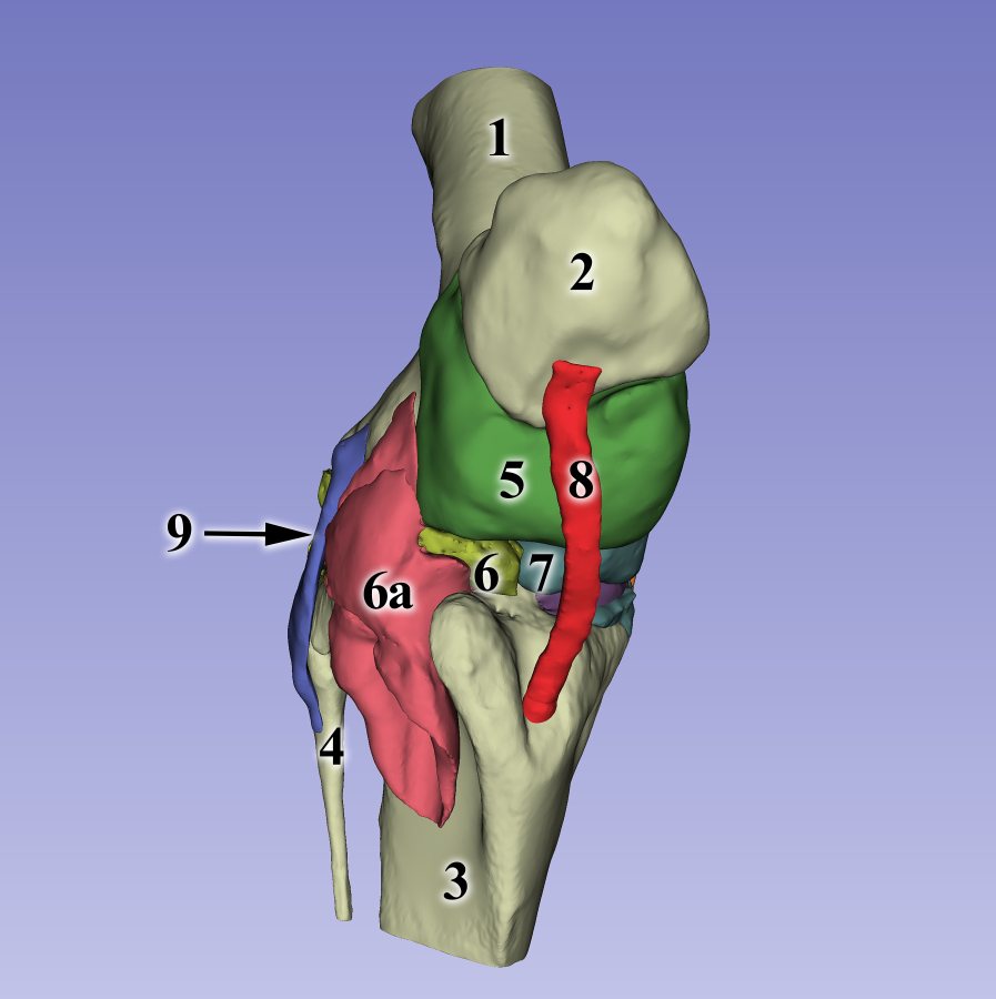
3D reconstruction of the equine stifle joint, anterior view
- femur
- patella
- tibia
- fibula
- femoropatellar joint (articulatio femoropatellaris)
- lateral femoral-tibial recess (recessus femorotibialis lateralis)
- medial femoral-tibial recess (recessus femorotibialis medialis)
- middle patellar ligament (lig. patellae intermedium)
- lateral collateral ligament (lig. collaterale laterale)
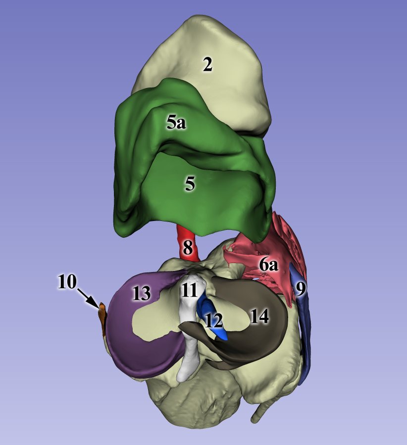
3D reconstruction of the equine stifle joint, posterior view, without the femur
- femoropatellar joint (articulatio femoropatellaris);
- a.recessus subextensorius
- middle patellar ligament (lig. patellae intermedium)
- lateral collateral ligament (lig. collaterale laterale)
- medial collateral ligament (lig. collaterale mediale)
- caudal cruciate ligament (lig. cruciatum caudale)
- cranial cruciate ligament (lig. cruciatum craniale)
- meniscus medialis
- meniscus lateralis
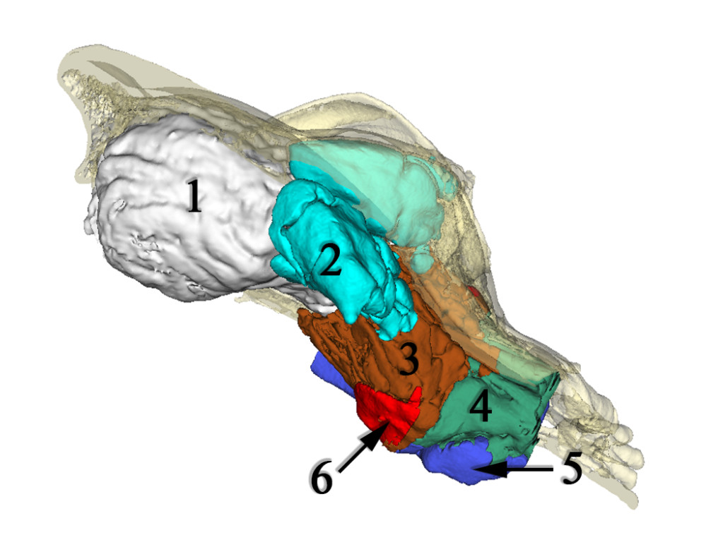
3D reconstruction of the canine skull
- neurocranium, endocast
- sinus frontalis
- cellulae ethmoidales
- ventral nasal conchae (concha nasi ventralis)
- ventral nasal meatus (meatus nasi ventralis)
- recessus maxillaris
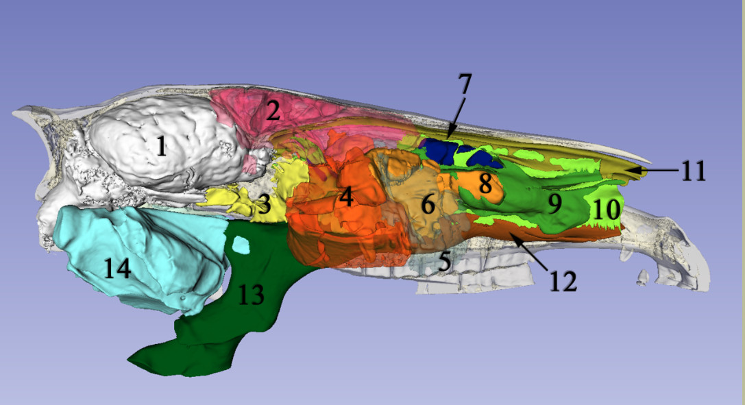
3D reconstruction of the equine skull
- neurocranium, endocast
- sinus frontalis
- sinus sphenoidalis
- sinus maxillaris caudalis
- sinus maxillaris cranialis
- bulla conchae ventralis
- sinus conchae dorsalis
- sinus conchae ventralis
- meatus nasi medius
- meatus nasi communis
- meatus nasi dorsalis
- meatus nasi ventralis
- pharynx
- diverticulum tubae auditivae
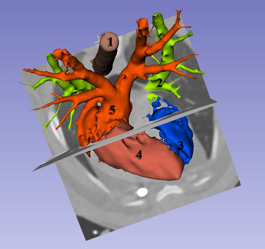
3D reconstruction of the swine heart
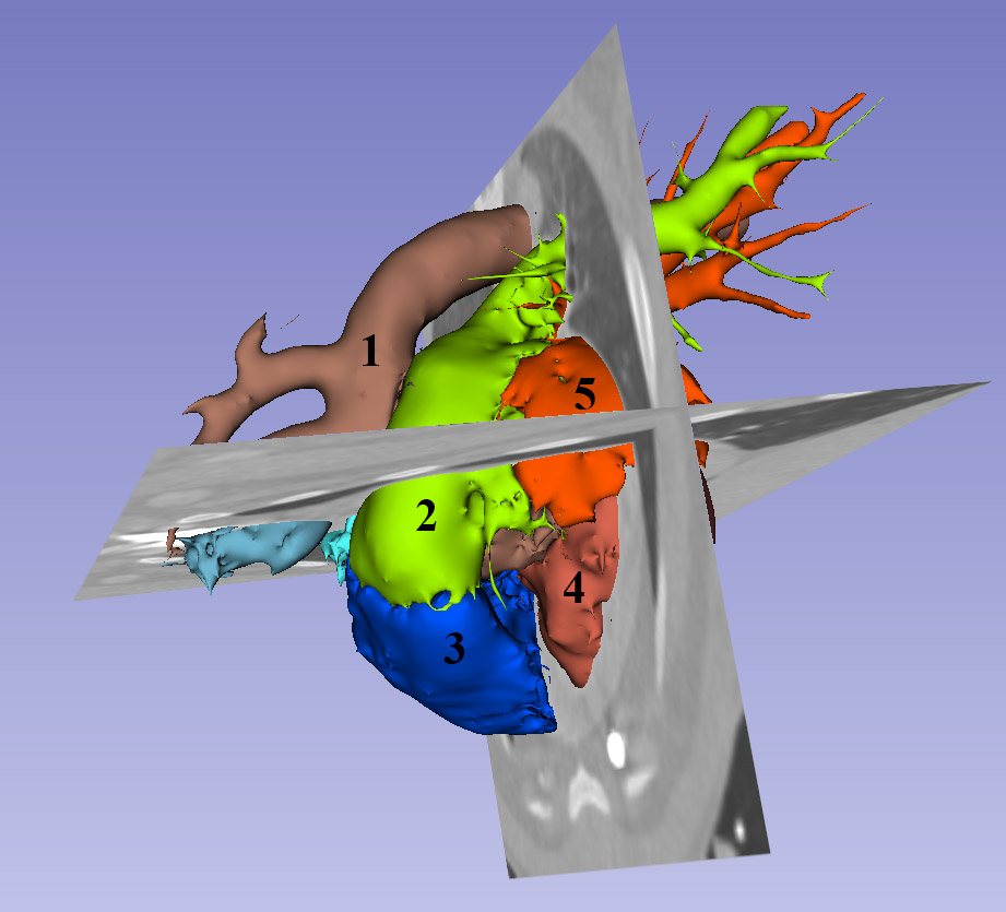
3D reconstruction of the swine heart
- aorta
- truncus pulmonalis
- right ventricle (ventriculus dexter)
- left ventricle (ventriculus sinister)
- left atrium (atrium sinister)
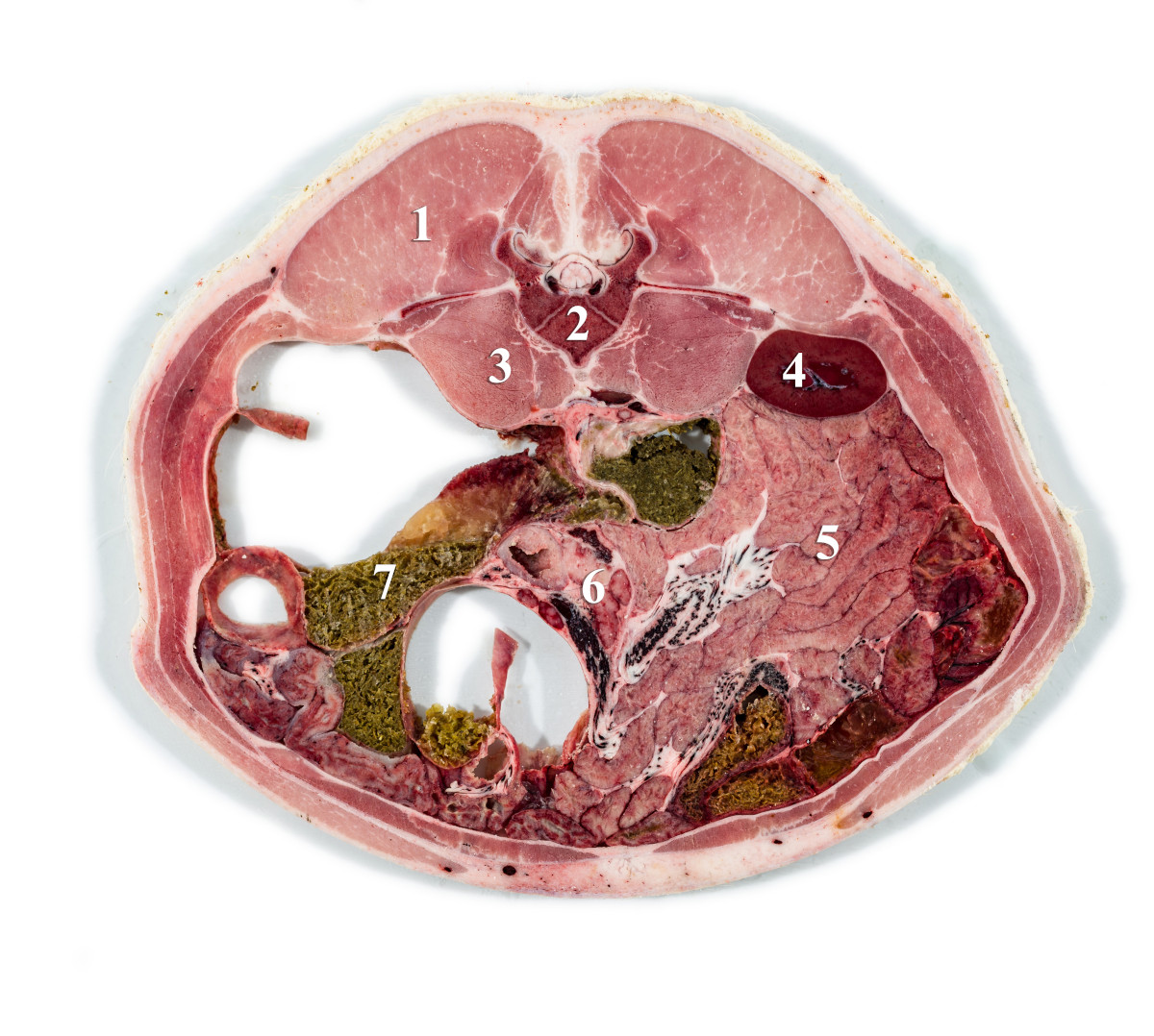
Cross-sectional image of the swine abdomen
- m. longissimus dorsi
- vertebra lumbalis
- m. psoas maior et minor
- right kidney (ren dexter)
- small intestines (jejunum)
- pancreas
- large intestines (colon)
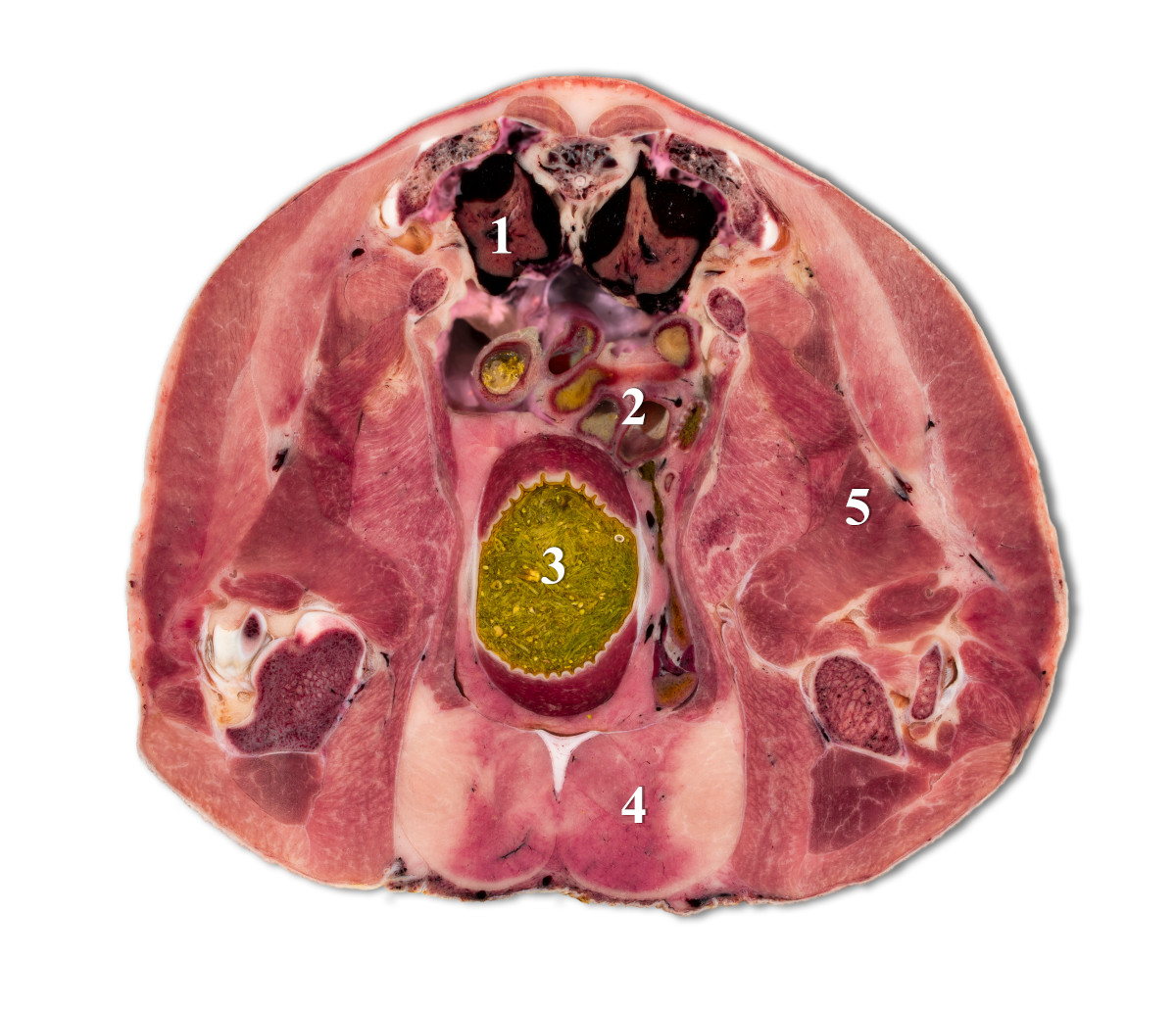
Cross-sectional image of the turkey abdomen
- left kidney (ren sinister)
- small intestines (jejunum)
- gizzard (ventriculus muscularis)
- pectoral muscles (m. pectoralis superficialis)
- thigh muscles (mm. femorales)